
Microtubule Definition
The microtubules in a cell’s cytoskeleton are microscopic hollow tubes made of the proteins alpha and beta tubulin. This cytoskeleton gives the cell shape and keeps its organelles in place by creating a network of protein filaments.
A microtubule measures about 24 nanometers thick, which makes them the largest structure in the cytoskeleton. Among their functions are cell movement, cell division, and transport of materials within the cell.
Microtubule Structure
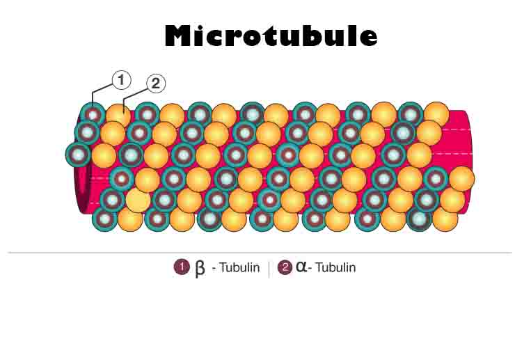
In microtubules, repeating protein structures, namely dimers of alpha and beta tubulin, are arranged into hollow cylinders. A dimer is a complex of two proteins. Protofilaments are formed when multiple units of alpha-tubulin and beta-tubulin bind together to form a chain, always alternately alpha and beta, called a dimer.
A microtubule is formed by arranging thirteen protofilaments in a cylindrical pattern. As dimers are added and removed, microtubules are constantly assembling and disassembling. Their structure is maintained despite the fact that the individual molecules themselves are constantly changing, and that is what is meant by dynamic equilibrium.
There are two ends of microtubules, one positively charged and one negatively charged. Microtubules are polar molecules. The beta subunits of a microtubule are always exposed on the positive end of the microtubule, while the alpha subunits are always exposed on the negative end. Polarity enables microtubules to assemble and function correctly in a specific manner.
Microtubules radiate outward from a microtubule organizing center (MTOC) in the center of animal cells called the centrosome. A nuclear envelope surrounds the nucleus of plants and fungi instead of centrosomes.
Function of Microtubules
Cell Movement
Flagella and cilia are composed of microtubules. An ilia is a small protuberance on the surface of a cell. Humans have them on the trachea, where they prevent mucus and dirt from entering the lungs. They are also found in the fallopian tubes of the female reproductive system, where they help move eggs from the ovary to the uterus. Cells move through flagella, which are tail-like appendages.
The flagella are also found in some bacteria and in human sperm. By contracting and expanding at opposite ends of the cell, microtubules allow whole cells to “crawl” or migrate.
Cell Division
As part of the mitotic spindle, also known as the spindle apparatus, microtubules play a key role. When eukaryotic cells undergo mitosis (cell division), this structure is formed. As chromosomes divide during cell division, the mitotic spindle organizes and separates them into two separate daughter cells. These include microtubules, MTOCs, and microtubule-associated proteins (MAPs).
There are three types of microtubules involved in mitosis: astral, polar, and kinetochore microtubules. Mitotic spindles are maintained by astral microtubules radiating from MTOCs to the cell membrane.
Chromosomes are separated by polar microtubules that intertwine between two MTOCs. (Microtubules are always polar; polar microtubules are just specifically called polar microtubules.) Kinetochore microtubules attach to chromosomes to help them separate.
Cell Transport
Microtubules are part of the cytoskeleton, which helps organelles move within a cell’s cytoplasm, which is its contents except for its nucleus. Additionally, they facilitate communication between different parts of the cell. Even though microtubules enable components of the cell to move, they also provide shape and structure to the cell.
Other Cytoskeletal Components
Microfilaments and intermediate filaments make up the rest of the eukaryotic cytoskeleton. At about 7 nm in diameter, microfilaments are smaller than microtubules. Their function is to aid in the division of cytoplasm during cell division, as well as to facilitate cytoplasmic streaming, which is the movement of cytosol (cell fluid) throughout the cell.
Microfilaments are larger than intermediate filaments, but microtubules are smaller. As well as providing structural support, they help give the cell its shape.
Related Biology Terms
- Microtubules are made up of tubulin, a protein.
- Except for the nucleus, cytoplasm is all the contents of a cell.
- Microtubules form the spindle apparatus during mitosis, which separates chromosomes.
- In the mitotic spindle, kinetochore proteins attach chromosomes to microtubules.